Retinal detachment | Causes & Help: Dr. med. Bányai explains
Retinal detachment (amotio retina) is a relatively rare condition affecting the retina. It is characterized by the inner layers of the retina detaching from their underlying nourishing layer (the retinal pigment epithelium). This detachment is usually caused by the natural aging process of the retina.
If the affected layer of the retina within the eye’s vitreous body is operated on early, the chances of recovery from a retinal detachment are very good. However, if a retinal detachment is treated too late or not at all, the detachment can lead to complete blindness.
Retinal detachment can be treated with laser eye surgery or with retinal surgery. Whether retinal surgery is needed depends on the stage of the retinal detachment. To arrange this, book an appointment with the ophthalmologist.
How is the retina structured?
The retina consists of several parts and lines the inner wall of the eye. The largest portion is the pars optica, which contains the photoreceptors—rods and cones. The point where the retina transitions into the optic nerve is called the blind spot.
The rods in the eye are primarily responsible for perceiving black and white. The cones, on the other hand, are responsible for perceiving colors. In addition, the cones are less sensitive to light, which is why a person often sees less clearly at night.
The nerve cells process the impulses they receive from rods and cones. These signals are then transmitted to the brain via the optic nerve, where the complete visual image is assembled.
What are the symptoms of retinal detachment?
Symptoms of retinal detachment – Retinal detachment usually manifests as various visual disturbances: patients see flashes of light, moving black dots and floaters. Spreading shadows can also occur.
If you notice even the slightest signs of retinal detachment in your eye, it is advisable to have your eye, retina, and vitreous body examined by an ophthalmologist as soon as possible.
The sooner a retinal detachment is detected and treated by an ophthalmologist, the better the chances of a full recovery. If left untreated or treated too late, however, a retinal detachment can lead to blindness in the affected eye.
How is a retinal detachment examined and diagnosed?
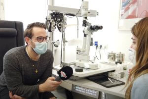
If your eyes show symptoms of a retinal detachment, you should see an ophthalmologist immediately. The ophthalmologist will perform an ophthalmoscopic examination of the vitreous body. This allows the inner surface of the vitreous, which is normally not visible from the outside, to be examined.
One advantage of an examination of the vitreous body is that it is painless and low-risk. It also allows a thorough inspection of the patient’s retina. The retina is examined for possible detachments, tears, or holes by the doctor illuminating the back of the eye or the vitreous body, enabling detection of any retinal changes.
Should a retinal detachment occur, the retina detaches from the choroid into the vitreous body. If the choroid separates from the retina, blood and other fluids can collect in the patient’s eye. Damage to the choroid and fluid accumulation should be avoided. If a clear assessment of the vitreous is not possible, ultrasound is a common alternative for detecting retinal detachment.
Retinal detachment: What are the risk factors and causes?
Die Netzhautablösung ist eine relativ seltene Krankheit. Die Ablösung der Netzhaut bzw. Retina betrifft statistisch nur einen von 10.000 Menschen. Das entspricht 0,01 Prozent. Dennoch können auch Löcher entstehen. Meist tritt die Netzhautablösung der Augen zwischen dem 45. und 65. Lebensjahr auf.
Besonders gefährdet sind Augen von stark Kurzsichtigen und an Diabetes Erkrankten. Lassen Sie sich daher immer wieder von einem Augenarzt untersuchen. Zusätzlich dazu ist die Netzhautablösung der Augen aber auch erblich bedingt. Am häufigsten wird die Netzhautablösung durch den ganz normalen Alterungsprozess des Auges ausgelöst:
Der aus einer gallertartigen Masse bestehende Glaskörper des Auges, schrumpft im Laufe der Jahre. Dadurch entstehen Löcher und Flüssigkeit tritt aus. Die rhegmatogene Netzhautablösung des Auges ist die Folge. Zur Vorsicht sollte immer ein Augenarzt bei Symptomen aufgesucht werden.
Welche Möglichkeiten der Behandlung gibt es bei einer Netzhautablösung?
Bislang gibt es keine medikamentöse Behandlung der Netzhautablösung. Je nach Stadium und dem individuellen Krankheitsverlauf der Netzhautablösung kommen aber folgende Möglichkeiten zur Behandlung der Augen infrage:
Die Laser-Operation der Augen
Je eher die Ablösung der Netzhaut entdeckt wird, desto unkomplizierter und risikoärmer ist auch die Behandlung der Netzhaut des Auges. Wenn die Ablösung der Netzhaut noch in einem sehr frühen Stadium ist, bzw. wenn nur ein Riss in der Netzhaut vorliegt, ist in der Regel das Augen lasern als Behandlung am betroffenen Auge möglich.
Der Laserstrahl bewirkt an der verletzten Stelle des Auges eine Entzündung, die eine Vernarbung im Gewebe der Netzhaut bzw. Retina hervorruft und so den Riss in der Netzhaut des betroffenen Auges behandelt und verschließt. Meist ist eine operative Behandlung des Risses in der Netzhaut des Auges durch Lasern ambulant möglich.
Die Operation der Augen
Oft wird die Ablösung und Bildung der Löcher der Netzhaut aber erst in einem Stadium entdeckt, indem eine Laser-Behandlung der Netzhaut bereits wirkungslos ist. In diesem Fall muss das Auge einer operativen Behandlung unterzogen werden. Die Methode hierfür hängt stark von der Art der Netzhautablösung und dem Stadium der Netzhaut-Erkrankung ab.
Ziel der Operation an der Netzhaut ist aber in jedem Fall, die Netzhaut der Augen zu fixieren und den eigentlichen Auslöser der Netzhautablösung zu beseitigen. Meist erfordert eine Operation der Augen bzw. der Netzhaut einen mehrtägigen stationären Aufenthalt.
Sollten Sie bei sich Anzeichen für eine Netzhautablösung oder die Entstehung von Löchern (Netzhautloch) bemerkt haben – oder wurde bei Ihnen bereits eine Ablösung der Netzhaut bzw. Retina diagnostiziert –, zögern Sie bitte nicht, uns zu kontaktieren.
In unserem Augenlaserzentrum in Karlsruhe und Augenlaserzentrum in Stuttgart stehen wir Ihnen mit langjähriger Erfahrung und moderner Diagnostik zur Seite. Wir beraten Sie individuell über geeignete Behandlungsmöglichkeiten und klären Sie umfassend über das Thema Netzhaut, Netzhautablösung und mögliche Therapien auf.
Alle Infos zum Augenlasern – direkt in Ihr E-Mail Postfach!
NEWSLETTER
Folge uns